2003 Imaging Workshop for Renal Researchers

Past Workshops


As part of the educational component of the NIH George M. O'Brien Center Award, the Indiana Center for Biological Microscopy has hosted several intravital workshops for renal researchers throughout the world.
The workshops have provided renal investigators with formal training and hands-on experience with intravital microscopy of the kidney.
-
• Microscopy: Understanding fundamental principles and what’s new
-
• Selecting a fluorescence microscopy system based on your specific needs
-
• Introduction to Volume Rendering
-
• Strategies of fluorescence labeling and sample preparation to maximize interpretation
Exercises emphasized hands-on experience with multiple instruments for digital image collection and analysis. Participants conducted studies with the following equipment:
-
• Bio-Rad MRC-1024 confocal/multiphoton microscope
-
• Zeiss 510-NLO-META confocal/multiphoton microscope
-
• Zeiss 510-UV confocal microscope
-
• Perkin-Elmer Ultraview High-speed confocal microscope
-
• Applied Precision Deltavision image deconvolution microscope system
-
• Pentium-based image processing workstations with Metamorph and Voxx rendering software
Laboratory Exercises Included:
-
• Alignment and adjustment of the light microscope for transillumination and epifluorescence
-
• Collection of digital images: balancing the tradeoffs according to experimental needs
-
• Evaluating and optimizing a fluorescence microscope system: measuring efficiency and noise characteristics
-
• Collection and analysis of 3-dimensional images: digital deconvolution, confocal microscopy and multi-photon microscopy
-
• Imaging dynamics of living cells and tissues in 2 and 3 dimensions
-
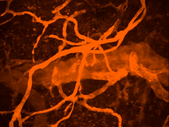
• Intravital microscopy: multiphoton microscopy of the kidney of a living rat
-
• Quantitative microscopy: ratiometric ion studies and evaluations of co-localization
Participants were encouraged to bring experimental materials to the workshop as center equipment and faculty will be available during evening hours for investigators to conduct pilot studies.
2003 Course Faculty & Lecturers:
-
Simon Atkinson - Indiana University School of Medicine
-
Robert Bacallao - Indiana University School of Medicine
-
Dennis Brown - Harvard Medical School
-
Kenneth Dunn - Indiana University School of Medicine
-
Marshall Montrose - Indiana University School of Medicine
-
Carrie Phillips - Indiana University School of Medicine
-
Kenneth Spring - NIH, National Heart, Lung and Blood Institute
-
George Tanner - Indiana University School of Medicine
-
Sam Wells - Vanderbilt Medical Center
Lecture Topics:
Laboratory Equipment: